Blog
Book your appointment with ChiroProActive online Book Chiropractor
X-ray or MRI, Imaging and symptoms
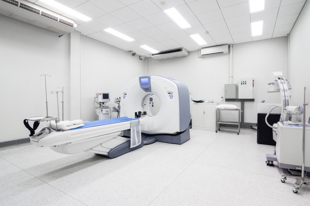
The use of imaging has increased significantly over recent years and so has the quality of imaging. There remains, however, significant strengths and weaknesses of different imaging types, as well as highly important concepts when we try to convert imaging findings into daily experiences. I will attempt to clarify because this is an important and persistent problem.
A bit of background first; as a regulated chiropractor in the UK, I and my colleagues graduate university with the training and competencies to take, read and report on x-ray images and with some training in the interpretation of MRI imaging (some other healthcare practitioners may not be aware of this). As such, some chiropractic clinics will have x-ray facilities on site. This, like so many things can be a double-edged sword.
Firstly, a clinic that has imaging facilities on site is more likely to use those facilities, not least because they need to raise funds to cover their costs. There are however regulations around the appropriateness of taking imaging. Nobody should be told they need to pay for a test they don't actually need. To be clear then, imaging taken with good reason and the right procedures is very safe and to be encouraged, but such reasons and procedures must be followed.
Secondly, we come to at least as important an issue, of what we do once an image has been taken. Far too often the imaging findings are used to explain symptoms, and this becomes the end of the conversation about an issue - something like, 'degenerative changes and disc bulge at L5-S1' in the low back. 'Now we know why you have low back pain'. The problem is that while this finding and statement appear to be valid on the surface, there are multiple systematic reviews of both x-ray and MRI and of multiple areas of the body (spine, hips, knees, shoulders as examples) (Brinjikji et al.), (Haonan et al.) that show very poor correlation between imaging findings and symptoms. To clarify, this means some people have degenerative changes and disc bulges with no symptoms and some people have minimal changes on imaging but lots of symptoms, and vice versa.
Ultimately degenerative changes with aging are normal. A 20-year-old looks younger than an 80-year-old and this is true on the surface and with imaging of the underlying structures. In conservative care (meaning non-invasive, non-surgical), unless we find that a person is unresponsive to care or we have good reason to suspect there is something otherwise underlying the problem (infection or fracture for example), we are generally best avoiding excess imaging and instead focussing on developing physical abilities and reducing pain (Tousignant-Laflamme et al.) with a combination of hands on treatments, progressive habit change and exercise progression towards meaningful goals and values. This leads to less pain and improved quality of life and positive changes with metabolic age (a subject for another time).
Please refer to previous blog articles on Specific Adaptation to Imposed Demand and Traffic Lights for additional related information.
Conclusion: If you find yourself in a situation where imaging has been taken and wear and tear has been identified, your experience and quality of life are still modifiable. If an issue is challenged appropriately the body will adapt positively. How you can use your body matters more than how it looks on a static image. This is what the chiropractic care including rehabilitation strategies we provide is dedicated to achieving.
References:
W. Brinjikji, P.H. Luetmer, B. Comstock, B.W. Bresnahan, L.E. Chen, R.A. Deyo, S. Halabi, J.A. Turner, A.L. Avins, K. James, J.T. Wald, D.F. Kallmes and J.G. Jarvik Systematic Literature Review of Imaging Features of Spinal Degeneration in Asymptomatic Populations. Am J Neuroradiol 2015, 36 (4) 811-816.
Haonan Fang, Xiaoyue Zhang, Junjie Wang, Xing Xing, Ziyuan Shen, Guoqi Cai. The relationship between MRI-detected hip abnormalities and hip pain in hip osteoarthritis: a systematic review. Rheumatology International (2024) 44:1887-1896.
Yannick Tousignant-Laflamme, Christian Longtin, Jean-Michel Brismee. EDITORIAL: How radiological findings can help or hinder patients' recovery in the rehabilitation management of patients with low back pain: what can clinicians do? Journal of Manual & Manipulative Therapy, 2017 VOL. 25, NO. 2, 63-65.
Contact Us
Have you checked our FAQs page? You may find the answer you are looking for there.
Please read our Privacy Policy
Book Chiropractor


Registered Address: Suite 411, Baltic Chambers, 50 Wellington Street, Glasgow, G2 6HJ
Registered in Scotland | Company Number: SC399248
Registered in Scotland | Company Number: SC399248
© 2013-2026 ChiroProActive Ltd | All Rights Reserved
Web Design Glasgow | valid xhtml | Privacy Policy | Terms of Service
Web Design Glasgow | valid xhtml | Privacy Policy | Terms of Service